. L'anatomia di animali domestici. Anatomia Veterinaria. Il peritoneo 443 a 30 cm.) sulla piccola colon. Si formano poi.s un angolo acuto, passa medialmente e in avanti lungo il colon di piccole dimensioni per la parte dorsale del hilus della milza, dove si mescola con il legamento sospensivo di quest'ultimo, e forma un incavo (Recessus lienalis) dietro il caucus saccus dello stomaco. Si passa ora lungo la hilus della milza ed è continuato per la maggiore curvatura dello stomaco dall'apparato gastro-cellule spleniche omento. È conveniente considerare la milza come essendo interca- affini nella parte sinistra del grea
1389 x 1799 px | 23,5 x 30,5 cm | 9,3 x 12 inches | 150dpi
Altre informazioni:
Questa foto è un'immagine di pubblico dominio, il che significa che il copyright è scaduto o che il titolare del copyright ha rinunciato a tale diritto. Alamy addebita un costo per l'accesso alla copia ad alta risoluzione dell'immagine.
Questa immagine potrebbe avere delle imperfezioni perché è storica o di reportage.
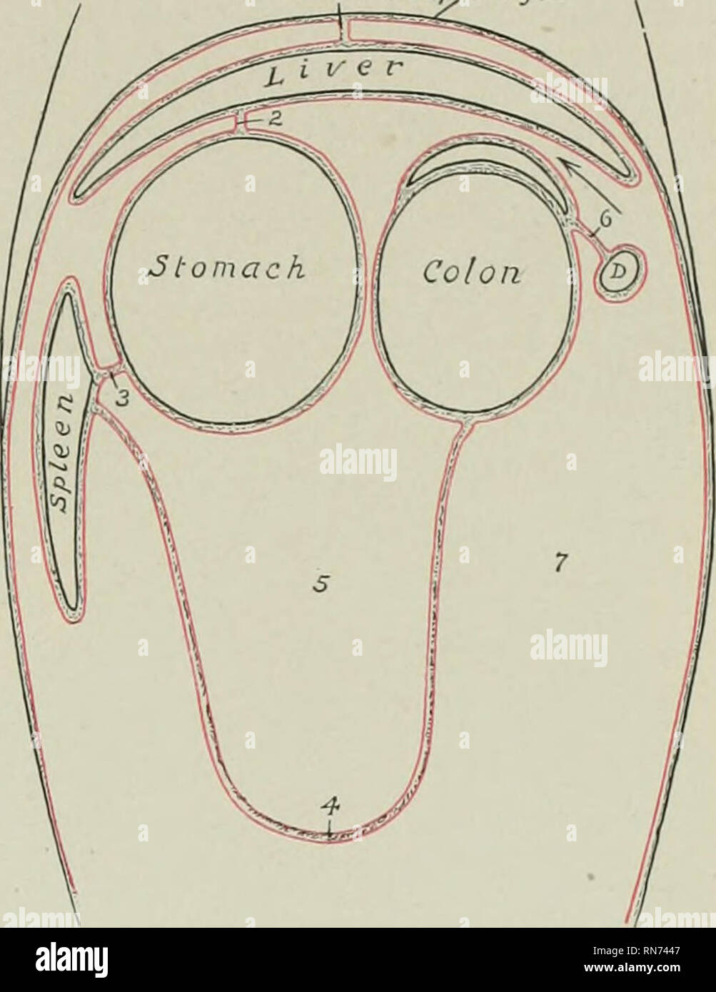
. The anatomy of the domestic animals. Veterinary anatomy. THE PERITONEUM 443 to 30 cm.) on the small colon. It then form.s an acute angle, passes medially and forward along the small colon to the dorsal part of the hilus of the spleen, where it blends with the suspensory ligament of the latter, and forms a recess (Recessus lienalis) behind the saccus caucus of the stomach. It now passes along the hilus of the spleen, and is continued to the greater curvature of the stomach by the gastro-splenic omentum. It is convenient to regard the spleen as being interca- lated in the left part of the greater omentum; on this basis the gastro-splenic omentum would be that part of the greater omentum which connects the hilus of the spleen with the greater curvature of the stomach. The greater omentum is relatively small in the horse, and is usually not visible when the abdo- ^ Diaphragm men is opened. It is generally folded up in the space between the visceral surface of the stomach and the in- testine.^ The lesser sac furnishes the perito- neal covering for: (1) the -isceral surface of the stomach and a small area of the first curve of the duodenum; (2) a large part of the dorsal surface of the pancreas and por- tal vein; (3) a small part of the visceral sur- face of the liver above the attachment of the lesser omentum and the portal fissure; (4) the posterior vena cava, from the level of the epi]>loic foramen to its passage through the diaphragm (in so far as it is not embedded); (5) the part of the parie- tal surface of the Uver between the right and middle divisions of the coronary liga- ment; (6) the corresponding part of the diaphragm, and the right part of the right crus of the same; (7) part of the anterior surface of the terminal part of the great colon, and the origin of the .small colon; (8) the left extremity of the pancreas (in- constant); (9) the spleen. We may now trace the perito- neum in a longitudinal direction, be- ginning in front. It is reflected from the