. Cunningham il libro di testo di anatomia. Anatomia. ondary coriali â Amnion cavità Amnion - Corpo-levetta Allantoic diverti- culum rimitive streak eurenteric cavità sul canale di entoderm sac extra-celom embrionale Decidua capsularis Decidua vera sono embrionali Fig. 75.âSchema di una sezione di una gravidanza utero dopo la formazione del Intervillous Spaces. parti. (1) Le parti che si estendono fra adiacenti spazi di sangue, il primario villi coriali. (2) Le parti che si trovano in con- tatto con il mesoderma del corion e che forma con il mesoderma del corion piastra. (3) Le parti che coprono la materna tissu
1592 x 1569 px | 27 x 26,6 cm | 10,6 x 10,5 inches | 150dpi
Altre informazioni:
Questa foto è un'immagine di pubblico dominio, il che significa che il copyright è scaduto o che il titolare del copyright ha rinunciato a tale diritto. Alamy addebita un costo per l'accesso alla copia ad alta risoluzione dell'immagine.
Questa immagine potrebbe avere delle imperfezioni perché è storica o di reportage.
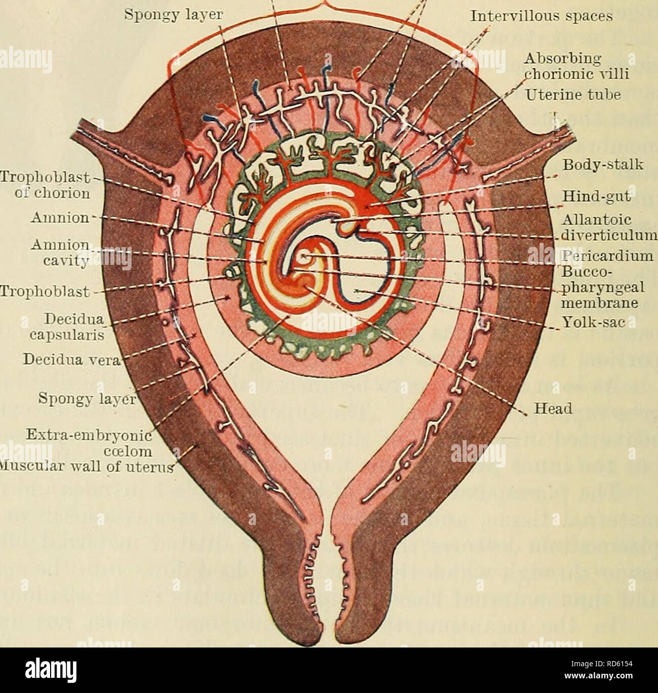
. Cunningham's Text-book of anatomy. Anatomy. ondary villus â Amnion cavity Amnion -Body-stalk Allantoic diverti- culum rimitive streak eurenteric canal Cavity of entoderm sac Extra-embryonic celom Decidua capsularis Decidua vera Embryonic are Fig. 75.âSchema of a Section of a Pregnant Uterus after the formation of the Intervillous Spaces. parts. (1) The parts which lie between adjacent blood spaces, the primary chorionic villi. (2) The parts which lie in con- tact with the mesoderm of the chorion, and which form with the mesoderm the chorion plate. (3) The parts which cover the maternal tissues and form the outer boun- daries of the blood spaces, the basal layer. The blood spaces themselves are called the in- tervillous spaces (Figs. 76, 79). After a time each primary villus differenti- ates into a cellular core and plasmodial periphery, and thereafter the villi are invaded by the mesoderm of the chorion and are thus converted into secondary villi (Fig. 76). The first-formed villi are non-vascular, but by the time the secondary villi have developed the um- bilical arteries have grown through the body-stalk (allantoic stalk) into the meso- derm of the chorion, and branches from them enter the meso- dermal cores of the villi, which thus be- come vascular. When the second- ary villi are fully developed each con- sists of a vascular mesodermal core con- tinuous with the mesoderm of the chorion Thp mpsn / 6.âSchema of a Frontal Section of a Pregnant Uterus at the vj uxi. j.ire luesu- period of the Formation of the Embryo. Xote extension of amnion dermal core IS Covered as contrasted with stage shown in Fi°- 75 Unchanged layer Maternal blood-vessels "i Placental area Spongy layer âV"^, , , "" "--^/â / Intervillous spaces Absorbing horionic villi Uterine tube Body-stalk Trophoblast-. Decidua capsularis Decidua v Spongy lay. Extra-embryon ecelom Muscular wall of uterus-'. Please note that these images are extracted from scanned page images tha