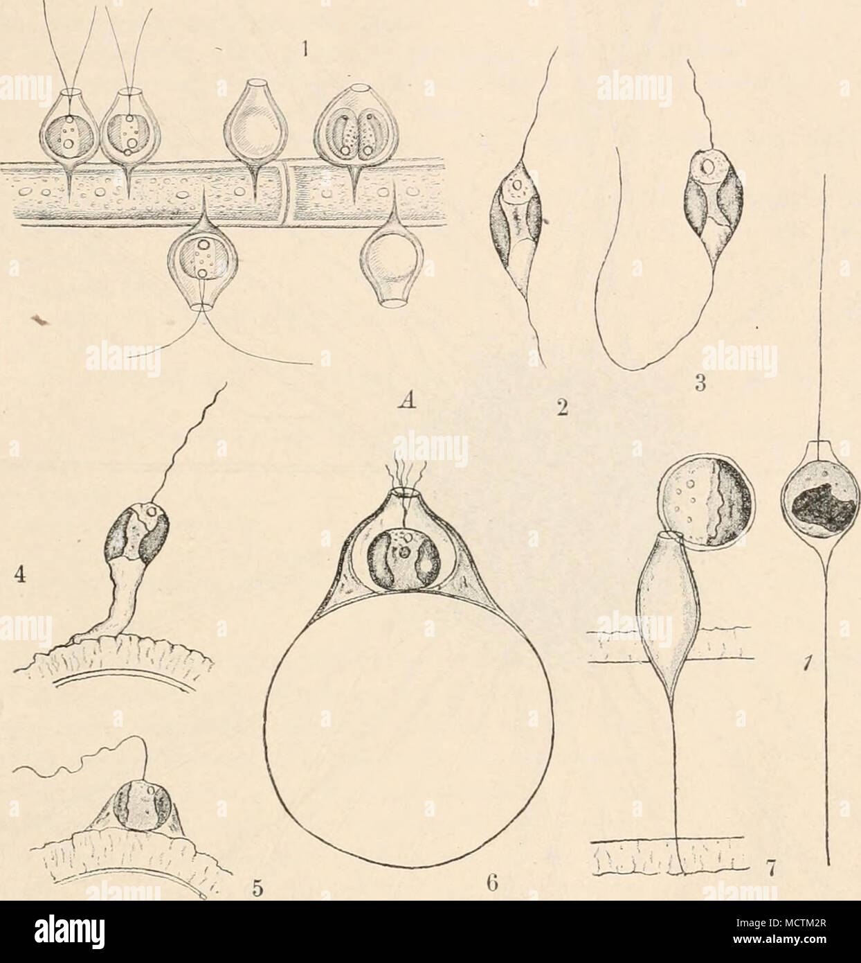
. Fig. 110. Un Chrysopyxis bipes Stein. 1 Zellen auf Cladophora. 2 und 3 freie Zellen, Entstehung des Sehwauz- fadens. 4 und 5 Herumwandern und Festsetzung auf einem Zygnemafaden. 6 Zelle mit vollständigem ßing von vorn gesellen. 7 Dauercyste an der Mündung des Gehäuses; dieses von der Seite gesehen. Vergr. ca. 800mal.- B Stylococcus aureus Chodat. 1 und 2 verschiedene Formen der Zellen. 3 und 4 Querteilung mit nachheriger Verschiebung. Vergr.? (1 nach Stein (1S7SJ; 2-7 nach Iwanoff (1899); b nach Chodat (1897).) Leucosinkörnchen und 1 contrattile (?) vacuolo. Kern centrale. Ernährung wohl nie
RMID:ID dell’immagine:MCTM2R
Detagli dell'immagine
Collaboratore:
The Bookworm Collection / Alamy Foto StockID dell’immagine:
MCTM2RDimensioni dei file:
14,3 MB (336,6 KB Download compresso)Liberatorie:
Modello - no | Proprietà - noMi occorre una liberatoria?Dimensioni:
2187 x 2285 px | 37 x 38,7 cm | 14,6 x 15,2 inches | 150dpiAltre informazioni:
Questa foto è un'immagine di pubblico dominio, il che significa che il copyright è scaduto o che il titolare del copyright ha rinunciato a tale diritto. Alamy addebita un costo per l'accesso alla copia ad alta risoluzione dell'immagine.
Questa immagine potrebbe avere delle imperfezioni perché è storica o di reportage.