. L'anatomia di animali domestici . Anatomia Veterinaria. 444 SISTEMA DIGESTIVO DEL BUE il csRcum e la parte terminale del grande colon. Da questi si passa sulla destra per il duodeno, che costituisce la terza parte della mesoduodenum. Sulla sinistra si copre una parte della superficie ventrale e il bordo laterale del rene sinistro, da cui passa alla base della milza, formando la strato ventrale del suspen- sory hgament di quest'ultimo. Dietro la parte terminale del grande colon è riflesso dalla parete addominale attorno alla grande arteria mesenterica per formare la grande mesenterica. Beh
1498 x 1667 px | 25,4 x 28,2 cm | 10 x 11,1 inches | 150dpi
Altre informazioni:
Questa foto è un'immagine di pubblico dominio, il che significa che il copyright è scaduto o che il titolare del copyright ha rinunciato a tale diritto. Alamy addebita un costo per l'accesso alla copia ad alta risoluzione dell'immagine.
Questa immagine potrebbe avere delle imperfezioni perché è storica o di reportage.
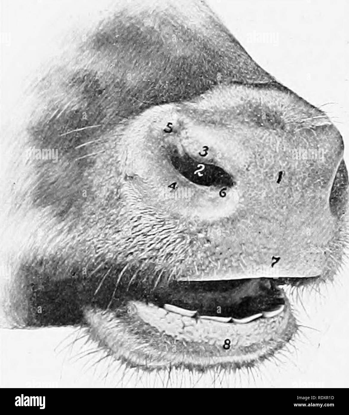
. The anatomy of the domestic animals . Veterinary anatomy. 444 DIGESTIVE SYSTEM OF THE OX the csRcum and the terminal part of the great colon. From these it passes on the right to the duodenum, forming the third part of the mesoduodenum. On the left it covers part of the ventral surface and the lateral border of the left kidney, from which it passes to the base of the spleen, forming the ventral layer of the suspen- sory hgament of the latter. Behind the terminal part of the great colon it is reflected from the abdominal wall around the great mesenteric artery to form the great mesentery. Behind this it is reflected almost transversely from the roof of the cavity and from the origin of the small colon on to the duodenum, forming the terminal part of the mesoduodenum. The line of origin of the colic mesentery begins on the medial part of the ventral surface of the left kidney, and extends to the sacral promontory, where the mesorectum begins. At the termination of the latter the peritoneum is reflected from the rectum on to the dorsal and lateral walls of the pelvic cavity. Below the rectum it forms the genital fold, and passes on to the dorsal surface of the bladder, covers its anterior part, and is reflected on to the body-wall laterally and ventrally, forming the lateral and middle ligaments of the bladder. In the female the broad ligaments of the uterus replace the genital fold, with which they are homologous. In the new-born foal certain folds are specially large. The falciform ligament of the liver extends to the umbilical opening, and contains in its free edge the large umbilical vein. The bladder—at this time an abdominal organ—has a ventral median fold (Plica umbilicalis media), which connects it and the urachus with the abdominal floor. This is flanked on either side by a fold (Plica umbilicalis lateralis), which also extends to the umbilicus, and contains the large umbilical artery. DIGESTIVE SYSTEM OF THE OX THE MOUTH The cavity of the mouth is short