. La diagnosi di malattie delle donne. Egli cellule sinciziali in-vaded gli spazi intermuscolari e le vene dell'utero fino al parametrio. Atrofia e necrosi degli elementi decidui e mus-circolari seguiti; I vasi sanguigni sono stati cambiati in lacunae del sangue. Nel confrontare il mio campione di mole idatiforme benigna con un DIAGI^OSIS DI HYDATIFORM MOLE 186 che ha subito alterazioni maligne, era visibile selezionare il confronto non solo uno di età simile, ma anche uno che aveva beenremoved insieme con l'utero, come era il mio. In questo modo eviti alcuni cambiamenti retrogradi e il disturbo di
1508 x 1657 px | 25,5 x 28,1 cm | 10,1 x 11 inches | 150dpi
Altre informazioni:
Questa foto è un'immagine di pubblico dominio, il che significa che il copyright è scaduto o che il titolare del copyright ha rinunciato a tale diritto. Alamy addebita un costo per l'accesso alla copia ad alta risoluzione dell'immagine.
Questa immagine potrebbe avere delle imperfezioni perché è storica o di reportage.
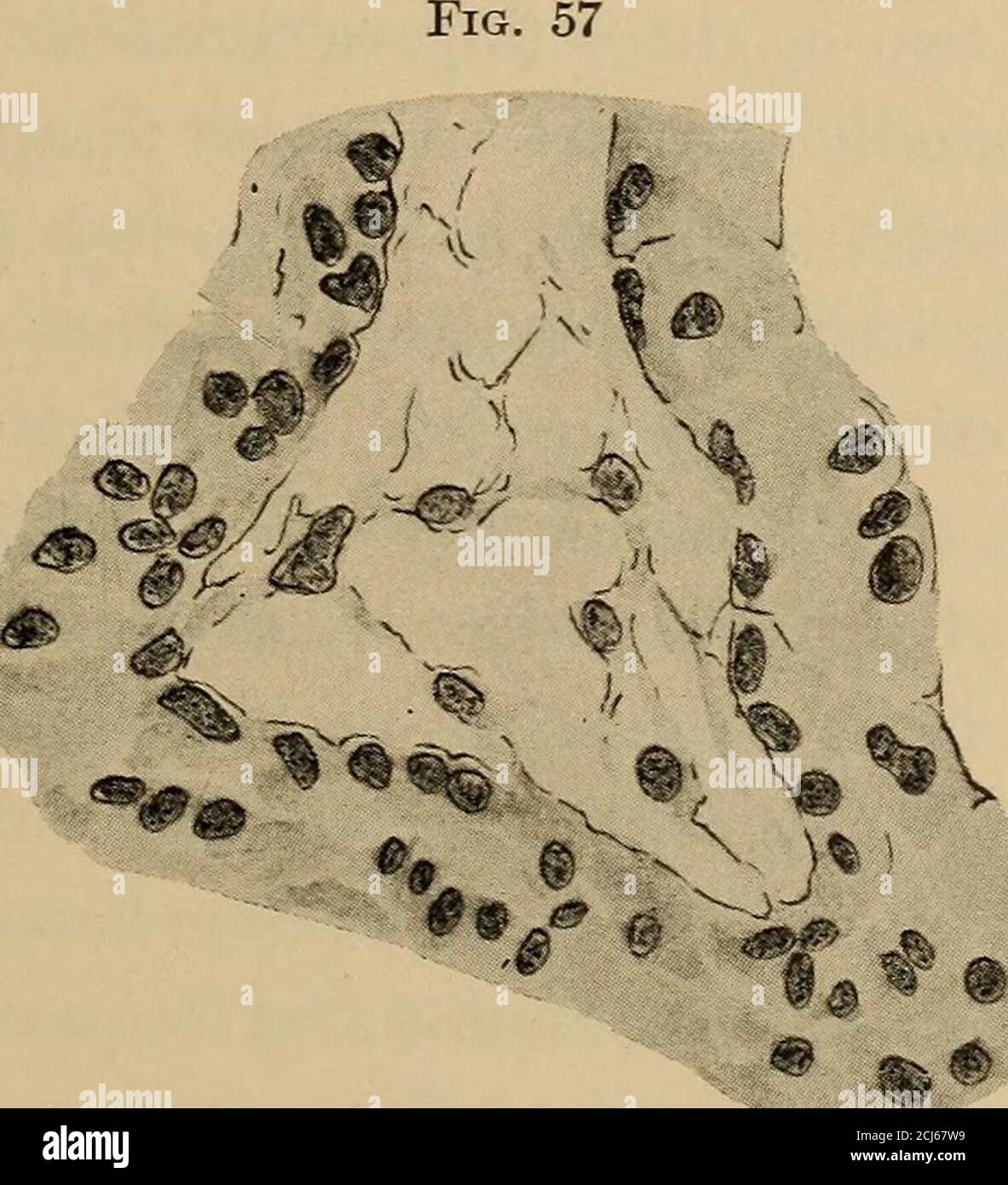
. The diagnosis of diseases of women . he syncytial cells in-vaded the intermuscular spaces and veins of the uterus as far asthe parametrium. Atrophy and necrosis of the decidual and mus-cular elements followed; bloodvessels were changed to blood lacunae.In comparing my specimen of benign hydatiform mole with one DIAGI^OSIS OF HYDATIFORM MOLE 186 having undergone malignant changes, it was advisible to select forcomparison not only one of similar age, but also one that had beenremoved together with the uterus, as was mine. In this way weavoid certain retrogressive changes and the disturbance of anatom-ical relations which would otherwise mislead. Two such caseshave been reported—one by Boten and Vassmer, the other byNeumann. In both these cases the essential variations from myown case appear to lie in the more marked proliferation of thesyncytium and Langhans cells and in their extended invasion ofthe uterine veins and musculature. While it is not to be expectedthat a benign mole may be recognized from a malignant mole by. Distal end of a chorionic villi, showing beginning degeneration of the stroma. the naked eye, yet it is worth while to observe that Pautz andothers have found in malignant moles that the villi rarely attainlarge size, are firm, and have a long, slender pedicle, giving to themole the appearance of soft-cooked rice. Ladinski, in a recent clinical review of deciduoma malignum, reported a case of hydatiform mole followed by malignant degenera-tion. He collected thirty-three similar cases, and concluded thatmalignant degeneration occurred most frequently in cases wheremole pregnancy terminated in the fourth month. It does notappear that the length of time a mole remains in utero has any influ-ence upon its disposition to become malignant. In twenty casesLadinski finds the average time of appearance of syncytiomamalignum is eight weeks after the mole has been expelled. 186 SPECIAL DIAGNOSIS CLINICAL DIAGNOSIS OF HYDATIFORM MOLE. The rate of growth of th